Diagnosis & Treatment Planning

Diagnostic imaging uses electromagnetic radiation, as well as other technologies, to generate extremely detailed images of your pet’s internal structures.

At Sydney Veterinary Emergency & Specialists, our team have access to the most advanced diagnostic tools and equipment available.

With our ability to perform diagnostics under one roof, we have the ability to expedite treatment for your pet.
In-House Lab & Pharmacy

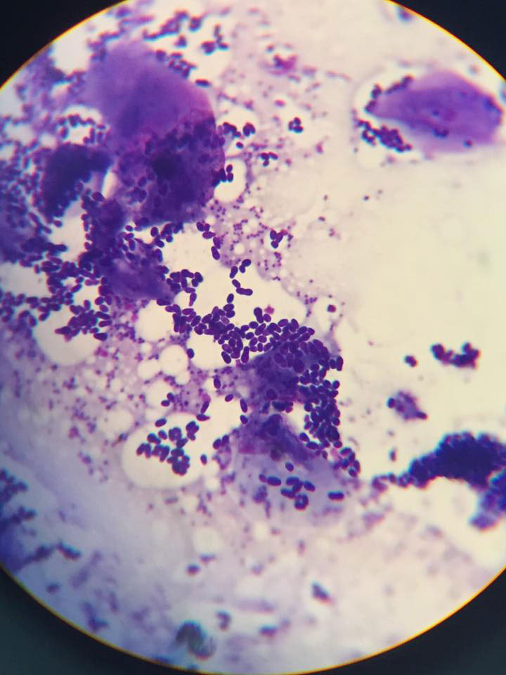
With our in-house laboratory, we can perform tests and get you results quickly, allowing us to assess your pet’s medical condition and start treatment planning. Our pharmacy features a wide range of medications and prescription diet foods, so we have immediate access to what your pet may need while in our care.

Diagnostic Services

An advanced yet non-invasive diagnostic machine, a CT scanner allows our specialists to produce very detailed cross-sectional images for the examination of your pet’s internal anatomy.

CT scans are used during the planning of surgery and disease diagnosis, as well as early-stage cancer detection and treatment.

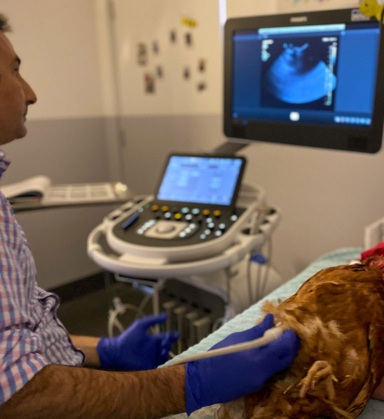
Ultrasound imaging produces picture of the inside of the body by exposing part of the body to high-frequency sound waves.

Ultrasound images are captured in real-time, so they can show blood as it flows through the blood vessels, as well as the structure of the body’s internal organs and their movement.

Also known as a digital x-ray, a radiograph looks inside your pet’s body and uncovers information that might not be evident from the outside.

Radiographs can be used to evaluate bones and organs and diagnose conditions including bladder stones, broken bones, chronic arthritis, spinal cord diseases and some tumours.

An endoscope is a flexible tube with an extremely small camera and light attached to the end.

An endoscopy inserts this tube directly into the body to take a closer, more detailed look at internal organs or tissue.

Endoscopes can be inserted with minimal invasiveness into openings of the body like the mouth or anus.

Fluoroscopy is a special type of x-ray imaging that allows us to see movement of body structures. For example, this type of imaging is very good for looking at dynamic bone instability, collapsing trachea, swallowing disorders, some cardiovascular disorders, and problems with urinary tract function.
Echocardiography is a special form of ultrasound that uses a top-of-the-line ultrasound unit to non-invasively ‘look inside the heart’. We are able to employ colour flow and contrast studies to safely diagnose heart conditions.
Magnetic resonance imaging (MRI) is used to help evaluate the soft tissues like the spinal cord, nerves, intervertebral discs, muscle and associated connective tissue, and the joints. Due to the excellent level of detail that MRI scans can offer, your specialist vet may prefer this method to diagnose brain and spinal cord issues in pets.